心血管类疾病模型,心血管疾病(CVD)是全球首要致死原因。为治愈疾病和降低一般人群中的疾病负担,开发能够改善乃至逆转疾病进程的新治疗方法十分必要。合适的动物模型为心血管疾病理论基础的探索、病因和发病机制的进一步明确及防范与治疗提供良好的研究平台。铂晋生物可提供以下几种心血管类疾病模型构建服务。
心肌梗死(myocardial infarction,MI)是一种严重的心血管疾病,由于冠状动脉血供急剧减少或中断,使心肌持续性缺血缺氧以致坏死,损害心功能且可能导致心律失常、休克以及心力衰竭等严重后果。
目前建立心肌梗死动物模型的方法有多种,包括:冠脉结扎法、药物法、栓塞法、左室电灼法、液氮冷冻法等。其中药物法主要通过舌下静脉注射垂体后叶素诱发血管痉挛促使心肌缺血梗死;后三种方法可直接形成冠脉闭塞,但对辅助设备要求高,操作难度大;冠状动脉结扎法操作成熟简便,梗阻部位明确易于判断,既符合心梗发生的实际病理过程,又便于开展实验研究,故主要采用此法建立心梗模型。
造模方法:结扎左冠状动脉前降支
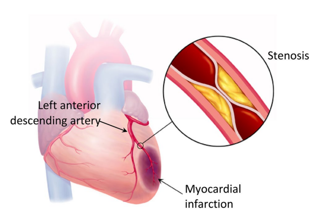
Fig. 1 Myocardial infarction caused from stenosis in left anterior descending (LAD) coronary artery.
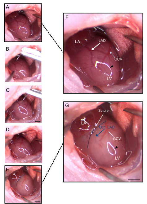
Fig. 2. Coronary Artery is Visible During Neonatal LAD Procedure.
模型评估及检测:
1、心电图看ST段的变化情况;
2、利用超声心动图进行心功能评价;
3、Masson染色观察心肌组织纤维化情况;
4、HE染色观察病理;
5、TTC染色观察梗死面积;
6、ELISA或qPCR检测TNF-α、IL-1β及IL-6等炎症因子。
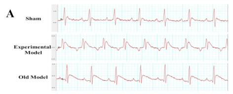
Fig. 3. Differences in statistical results between groups.(A), a higher ST-segment elevation (****p < 0.0001, *p = 0.0274).
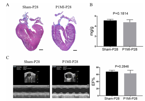
Fig. 4. P1 mice hearts show nearly full regeneration while P7 mice hearts show little regeneration after LAD ligation.(A)Representative images of Masson’s trichrome staining of P28 mice heart after P1 sham-operated or P1 MI. Hearts were longitudinally cut. Scale bar: 0.5 mm. (B)Heart weight to body weight ratio at P28. (C) Representative images of M-mode echocardiography and EF values at P28, n=5 for each group.
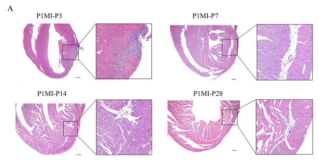
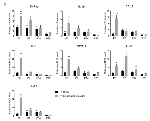
Fig. 5. Time‐course of the acute inflammatory response in mouse hearts after P1 MI. (A) Hematoxylin and eosin (HE) staining of P3, P7, P14 and P28 hearts after P1 MI.Representative images of low and high magnification views are shown. Scale bars, 200 µm. (B) qPCR assays of the expression of inflammatory markers (TNF-α, IL-1β, Ccl2, IL-6, Cxcl1, IL-17 and IL-23) in P3, P7, P14 and P28 heart tissues after P1 MI, n=5 per group, P1-Sham P28 group was set as control, *P<0.05, **P<0.01 compared with P1-Sham group at the same time point.
了解更多模型信息与服务细节,欢迎来电咨询。
服务热线:020-84781679