大脑中动脉闭塞(MCAO)模型,缺血性脑梗死具有高发病率、高死亡率、高致残率的特点。小鼠的脑血管解剖特点与人类较为相似,故广泛应用于缺血性脑梗死的研究当中。大脑中动脉闭塞(Middle Cerebral Artery Occlusion,MCAO)模型是指通过手术的方式,用特制线栓对大脑中动脉进行封堵形成短暂或永久性的血流阻断,使该动脉区域出现脑缺血状态。由于脑卒中患者中最常见的栓塞发生在大脑中动脉(MCA)区域,所以MCAO模型也作为模拟人类最常见的栓塞性局部缺血中风亚型而被广泛使用。
造模方法:游离左侧颈总、颈外和颈内动脉,结扎颈外动脉并离断,血管夹暂时夹闭颈总动脉和颈内动脉。线栓从颈外动脉近心端的切口进入颈内静脉,沿颈内静脉走行至大脑中动脉进行闭塞。

Fig. 1. Scheme of the occlusion of the middle cerebral artery using silicon-coated intraluminal monofilament. A. Simplified scheme of mouse brain and cerebral arteries showing successive sutures and clip to prepare the introduction of silicon-coated monofilament. B. The position of monofilament through the circle of Willis is represented. The monofilament is introduced into ICA via ECA to occlude the base of the MCA. ACA, anterior cerebral artery; BA, basilar artery; CCA, common carotid artery; C. Willis, Circle of Willis; ECA, external carotid artery; ICA, internal carotid artery; MCA, middle cerebral artery; PCA, posterior communicating artery; PPA, pterygopalatine artery(5).
模型评估及检测:
1、根据动脉梗塞的时间,会导致不同的运动和行为缺陷。一般用longa五分制评分法进行行为学评分。
0分:正常,无神经功能缺损;
1分:左侧前爪不能完全伸展,轻度神经功能缺损;
2分:行走时,大鼠向瘫痪侧转圈,中度神经功能缺损;
3分:行走时,大鼠身体向瘫痪侧倾倒,重度神经功能缺损;
4分:不能自发行走,有意识丧失。
缺血24h后进行评分,分值越高,说明动物行为障碍越严重,1-3分造模成功。
2、病理检测:大脑中动脉闭塞术后24h取材,进行TTC染色。
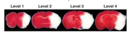
Fig. 2. Representative images of TTC-stained brain slices (coronal level 1-4) after 24 h of permanent MCAO. In living tissue TTC is enzymatically reduced by dehydrogenases to 1,3,5-triphenylformazan (TPF), which is red in color, while in necrotic areas it remains white due to absence of such enzymatic activity. Therefore, the area of infarction can be identified by its white color due to lack of conversion of TTC to TPF. Note: TTC is somewhat heat and light unstable so protect stained sections from heat and light as much as possible(6).
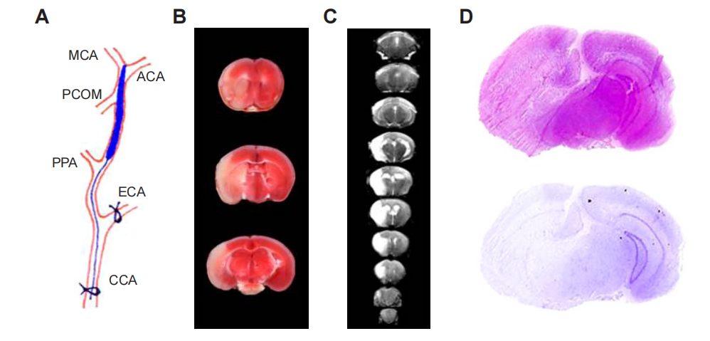
Fig.3. Scheme of an intraluminal suture MCAo model and different methods for determining infarct volume.
Notes: (A) Diagram of MCAo. (B) Representative of 2,3,5-triphenyl tetrazolium chloride staining of three consecutive coronal brain sections after transient MCAo. (C) Serial coronal T2-weighted gradient echo magnetic resonance images after transient MCAo. (D) Representative hematoxylin and eosin (top) and Nissl staining (bottom) of coronal brain sections after transient MCAo.(7)
了解更多模型信息与服务细节,欢迎来电咨询。
服务热线:020-84781679